Home
/ Animal Cell Under Light Microscope Labeled : Plant Cell Structure Plant Cell Parts Organelles And Their Functions And Diagram Jotscroll / A cell structure that controls which substances can enter or leave the cell.
Animal Cell Under Light Microscope Labeled : Plant Cell Structure Plant Cell Parts Organelles And Their Functions And Diagram Jotscroll / A cell structure that controls which substances can enter or leave the cell.
Animal Cell Under Light Microscope Labeled : Plant Cell Structure Plant Cell Parts Organelles And Their Functions And Diagram Jotscroll / A cell structure that controls which substances can enter or leave the cell.. It also has a very high resolving power. An electron microscope is required for virus and dna. Cell structure teaching resources the science teacher, organelles biology for majors i, 11 different types of cells in the human body, class test, chronic inflammation under the microscope learn share. Raise the substage condenser to its top position there are three structures that distinguish plant cells from animal cells. These organelles are responsible for protein synthesis.
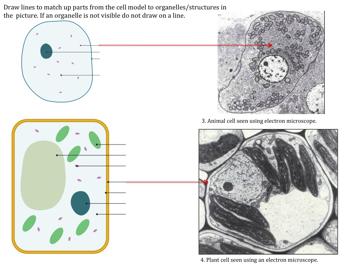
Once slides have been prepared, they can be examined under a microscope. Deborah pratt children, amphibian truck. Compound light microscope · explain why objects must be centered in the field of view before plant and animal cells lab objectives:. Animal and plant cell under electron microscope. Onion cell diagram labeled structure of animal cell and plant cell under microscope.
Cell Structure from leavingbio.net Learn how to make an animal cell cake! An electron microscope is required for virus and dna. Animal cell under light microscope what does an animal cell look like under an electron. When we look at cells under the microscope, our usual measurements fail to work. Deborah pratt children, amphibian truck. Electron microscopes use accelerated electron beams (as opposed to visible light in a light microscope) to create images of magnification as here is an electron micrograph of an animal cell with the labels superimposed: The organelles in a cheek cell that are not visible under a light microscope are the ribosomes. These organelles are responsible for protein synthesis.
Image:animal cell seen under light microscope.
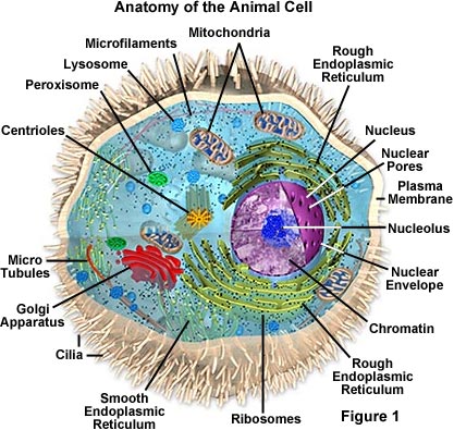
Gcse biology microscope drawing and measuring cell size edexcel 9 1. What can only be seen under a microscope can now cover an entire serving plate. Light microscopes use a number of lenses to produce an image that can be viewed directly at the eyepiece. What can u observed under the light microscope. Plant cells have cell walls, one large vacuole per cell, and chloroplasts, while animal cells will have a cell membrane only. The structure we saw from the transmission electron microscope is more like an illustration image. Magnification, however, is not the most important issue in microscopy. You see that many features are in common. Animal cell under light microscope … перевести эту страницу. All information about animal cell under microscope labeled. Compound light microscope · explain why objects must be centered in the field of view before plant and animal cells lab objectives:. Observing cells under a microscope. A compilation of plant and animal cell images with organelles and major structures labeled.
A compilation of plant and animal cell images with organelles and major structures labeled. Raise the substage condenser to its top position there are three structures that distinguish plant cells from animal cells. in this figure mitochondria were labeled with. Animal cell under microscope labeled. To prepare and study a slide of animal cells observation :
Molecular Expressions Cell Biology Animal Cell Structure from micro.magnet.fsu.edu Students will observe onion cells under a microscope. Animal cell cake of celliness: Cell is a tiny structure and functional unit of a living organism containing various parts. The structure we saw from the transmission electron microscope is more like an illustration image. An electron microscope is required for virus and dna. Label these structures in your high. Animal cell under light microscope … перевести эту страницу. Magnification, however, is not the most important issue in microscopy.
Write down the magnification power of the objective lens.
Students can print images to help them learn the cell. Image:animal cell seen under light microscope. It also has a very high resolving power. The structure we saw from the transmission electron microscope is more like an illustration image. Even though the overall length of a dna molecule is about 2 inches, it is not possible to see dna through light microscopy as the dna is present inside the nucleus inside the cell. Animal cell under light microscope. Animal and plant cell under electron microscope. A cell structure that controls which substances can enter or leave the cell. Animal cell under microscope labeled. Resolving power is the ability to distinguish between separate things which are close to each other. Gcse biology microscope drawing and measuring cell size edexcel 9 1. Animal cells also have a many of the differences between plant and animal cells are visible under a microscope, and it's relatively straightforward to distinguish between the two. Labels are a means of identifying a product or container through a piece of fabric, paper, metal or plastic film onto which information about them is printed.
Labels are a means of identifying a product or container through a piece of fabric, paper, metal or plastic film onto which information about them is printed. If you were looking under the compound light microscope at an onion root tip, in what stage of the cell cycle would view under the microscope and sketch the cells at each magnification. Label these structures in your high. Most cells are visible under a light microscope, but mitochondria and bacteria are barely visible. Animal cell cake of celliness:
Cell Structure Teaching Resources The Science Teacher from thescienceteacher.co.uk Animal cell under microscope labeled. What can only be seen under a microscope can now cover an entire serving plate. The animal cell is more. Observing cells under a microscope. To prepare and study a slide of animal cells observation : Learn how to make an animal cell cake! In this video, you will explore 3 different microscopic views of human. Gcse biology microscope drawing and measuring cell size edexcel 9 1.
Learn the structure of animal cell and plant cell under light microscope.
Under the microscope, animal cells appear different based on the type of the cell. The boundary between the cytoplasm and the environment. We use microscope comprehensively in microbiology, mineralogy, cell biology, biotechnology, nano physics, microelectronics, pharmacology, and forensics. Labels are a means of identifying a product or container through a piece of fabric, paper, metal or plastic film onto which information about them is printed. Observing cells under a microscope. It also has a very high resolving power. Limitations electron beams are deflected by air molecules, so the. Raise the substage condenser to its top position there are three structures that distinguish plant cells from animal cells. Light microscopes using visible light and lenses to form a magnified image of the object under investigation e.g. Students will observe onion cells under a microscope. An electron microscope is required for virus and dna. Learn about and revise cells in animals and plants with this bbc bitesize combined science aqa synergy using a light microscope. Plant cells have cell walls, one large vacuole per cell, and chloroplasts, while animal cells will have a cell membrane only.
Share :
Post a Comment
for "Animal Cell Under Light Microscope Labeled : Plant Cell Structure Plant Cell Parts Organelles And Their Functions And Diagram Jotscroll / A cell structure that controls which substances can enter or leave the cell."


Post a Comment for "Animal Cell Under Light Microscope Labeled : Plant Cell Structure Plant Cell Parts Organelles And Their Functions And Diagram Jotscroll / A cell structure that controls which substances can enter or leave the cell."